Fig. 1
From: Molecular and clinicopathologic characteristics of CNS embryonal tumors with BRD4::LEUTX fusion
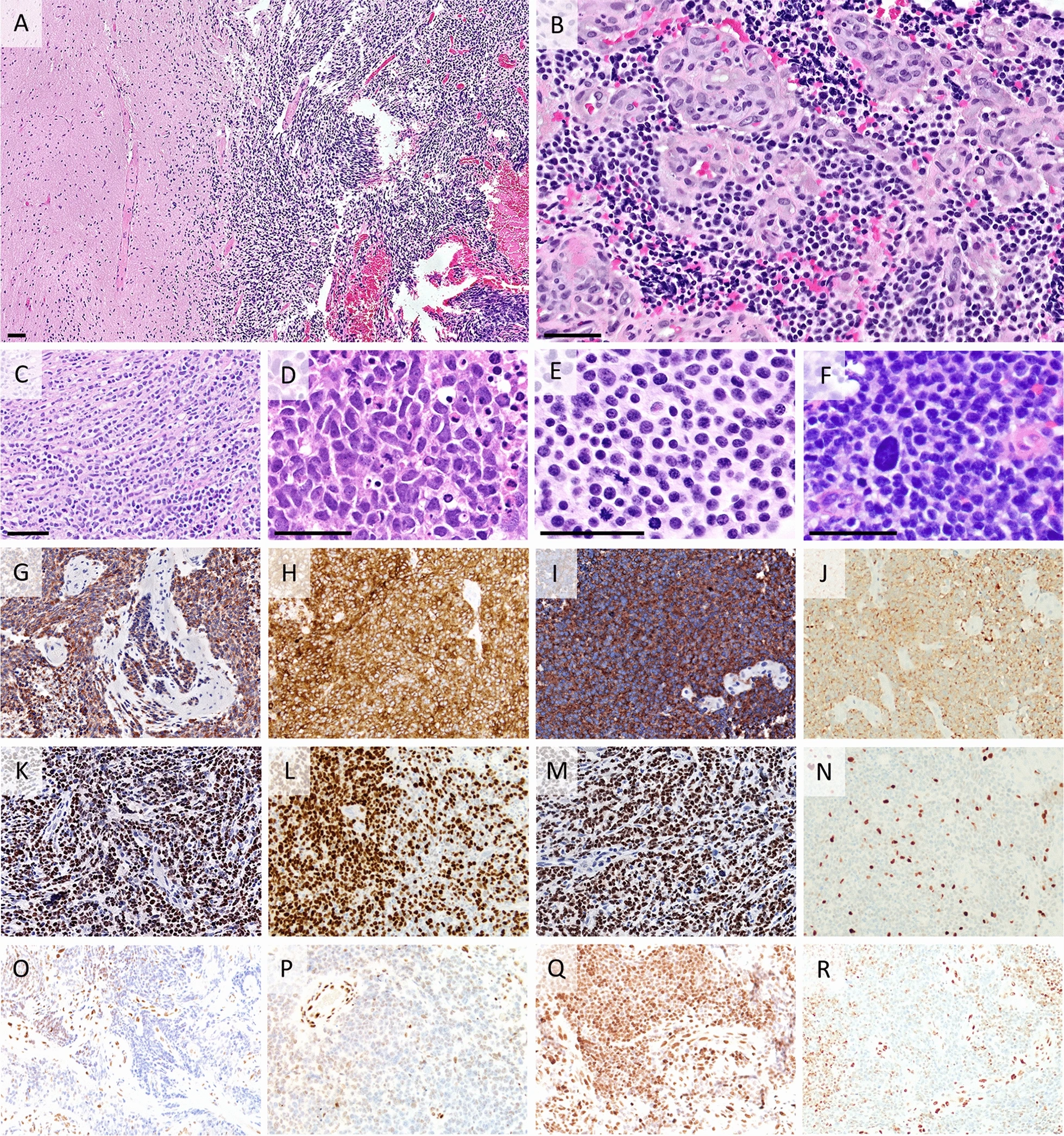
Histology and Immunohistochemistry. All scale bars measure approximately 50 µm. Immunostains were originally photographed at 20 × 10 = 200X magnification, with some images resized slightly to fit within the figure. A Case #3 was relatively well-circumscribed from adjacent nervous tissue. B Case #1 showed small monomorphic cells with round hyperchromatic nuclei and scant cytoplasm. Abundant proliferated vessels with particularly thick walls were seen at least focally in all cases. C Case #1: prominent cell streaming pattern. D Case #2 featured more irregularly shaped nuclei. E All cases had prominent mitotic activity as seen in case #3. F Case #4: An enlarged atypical nucleus stands out against small, regular nuclei. G–J Diffuse positivity of immunohistochemistry (IHC) for synaptophysin in cases #1 (G), #2 (H), #3 (I) and #4 (J). K–N. Expression of OLIG2 positivity seen in all cases, at variable levels. K Case #1 showed strong and diffuse positivity. L. Most cells were positive in case #2. M Case #3 displayed strong and diffuse positivity. N Case #4 showed scant OLIG2 positive cells. O–R H3K27me3 IHC for cases #1 (O), #2 (P), #3 (Q) and #4 (R). Q Case #3 showed retained expression. R Case #4 showed loss of expression in tumors cells with a mosaic pattern