Fig. 2

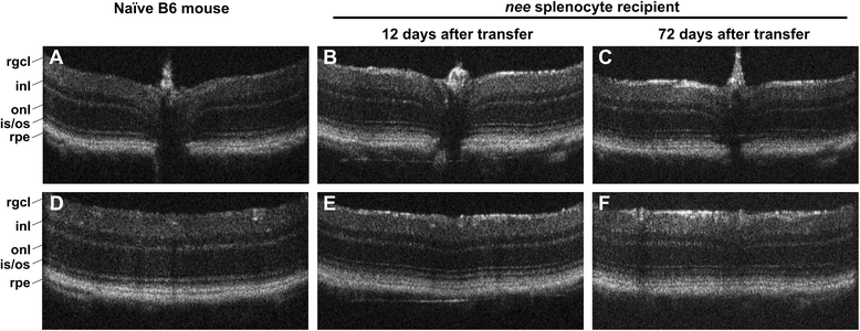
Optical coherence tomography. Retinal OCT imaging demonstrating a normal retinal architecture in the peripapillary (a, b, c) and peripheral regions (d, e, f) in both age-matched B6 naïve mice and a representative nee splenocyte recipient 12 and 72 days after transfer. No evidence for acute infiltration or retinal detachments was identified through in vivo imaging. A thinning of the retinal ganglion cell layer (rgcl, including the nerve fiber layer) was not yet evident 72 days after transfer, even though at that time a slight loss of RGC is observed using histochemical approaches. However an increased reflectivity (b, c, e, f) in the rgcl was noted when nee-transfer animals and naïve mice are compared (inl: inner nuclear layer; onl: outer nuclear layer; is/os: inner and outer photoreceptor cell segments; rpe: retinal pigment epithelium)