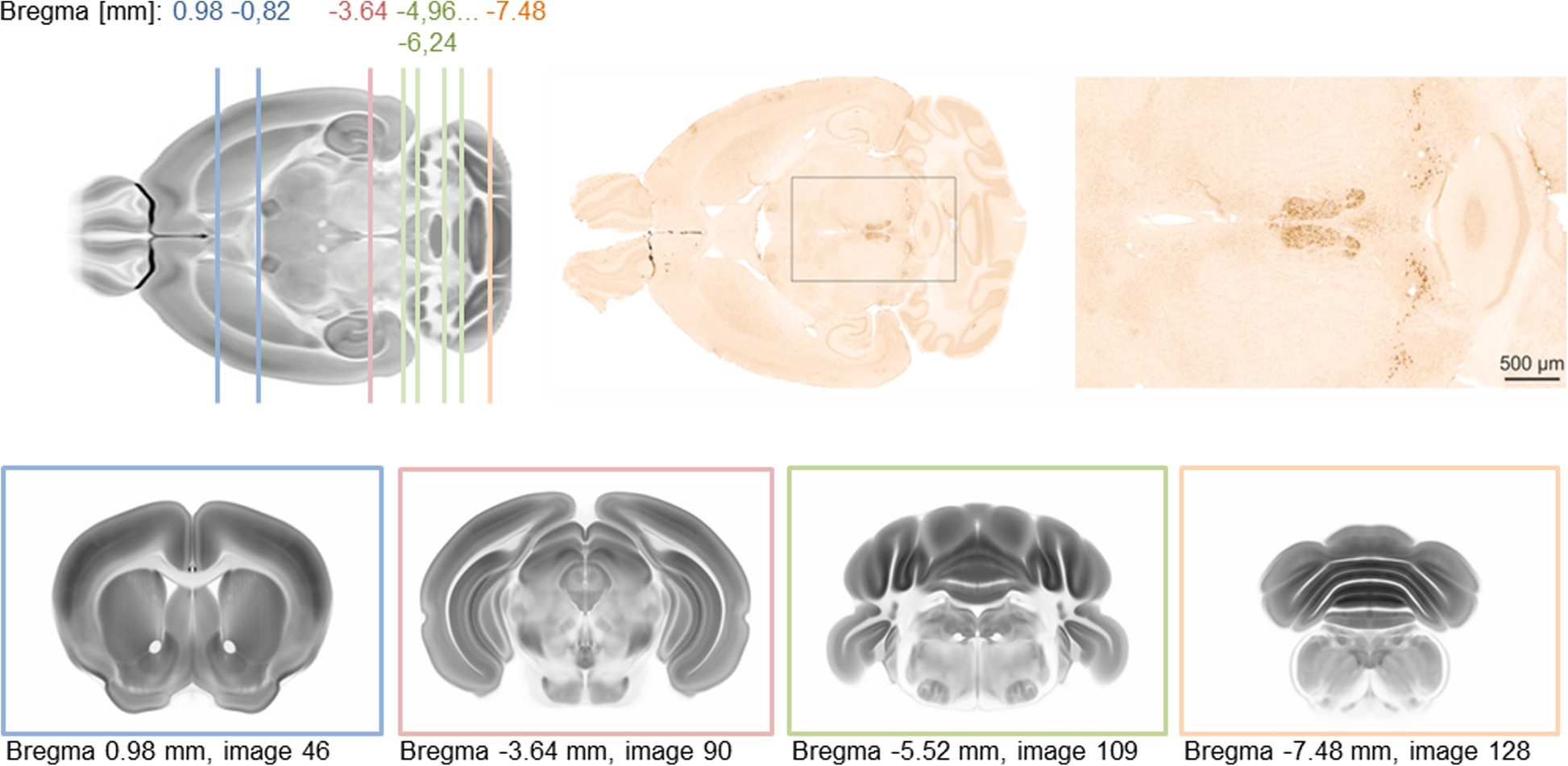
Fig. 1

Immunohistochemistry for huntingtin (HTT) on mouse horizontal brain sections (top, middle and right) and schematic presentation of coronal cutting levels with high abundance of strongly HTT immunoreactive neurons. Note the enrichment of HTT immunoreactivity in caudal brain structures. The anatomical templates used to illustrate cutting levels are shape and signal intensity averages from the Allen Mouse Brain Connectivity Atlas [60]