Fig. 5
From: Exosomes taken up by neurons hijack the endosomal pathway to spread to interconnected neurons

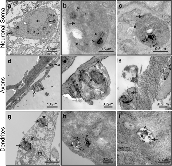
Hijacking of endogenous endosomes revealed by electron microscopy. Hippocampal neurons in microfluidic devices in Ch1 according to model 2 were treated with exosomes isolated from mouse brains and labeled with FM1–43FX, a fluorescent dye that reacts with diaminobenzidine (DAB) to form insoluble dark precipitates that are visualized by electron microscopy. a Electron microscopy image of a neuronal soma in Ch1, showing that the majority of endosomes contain DAB-positive exosomes of exogenous origin (black arrowheads). A few endosomes are DAB-negative (white arrowheads). b High magnification of endosomes located in the neuronal soma, showing a mixture of exogenous DAB-positive exosomes (black arrowheads) and endogenous DAB-negative intraluminal nanovesicles (black arrows). c Somatic endosome showing engulfment and fusion with a smaller endosome containing DAB-positive exosomes (black arrowhead “a”). An exogenous DAB-positive exosome can be seen close to the fused endosome (black arrowhead “b”). Endogenous DAB-negative intraluminal nanovesicles are also visualized (black arrows). d-f Endosomes found in axons. (d) Low magnification of axons transporting a small DAB-positive endosome (black arrowhead “a”) and a large endosome in front of it (black arrowhead “b”). e High magnification of a large axonal endosome containing a mixture of DAB-positive (black arrowheads) and DAB-negative vesicles (black arrows). f Axonal termini showing endosome fusion with the plasma membrane (indicated with “!”) during exosome release and potential residue of the back-fusion with the limiting membrane of the endosome (*). Exogenous DAB-positive (black arrowheads) and endogenous DAB-negative exosomes (black arrows). g-i Endosomes found in dendrites. g Low magnification of a dendrite demonstrating the presence of endosomes carrying both DAB-positive exosomes (black arrowheads) and DAB-negative intraluminal nanovesicles (black arrows). h and i High magnification of dendritic endosomes carrying DAB-positive exosomes less than 100 nm diameter (black arrowheads) together with DAB-negative vesicles of a similar size (black arrows). m, mitochondria; n, nucleus