Figure 1

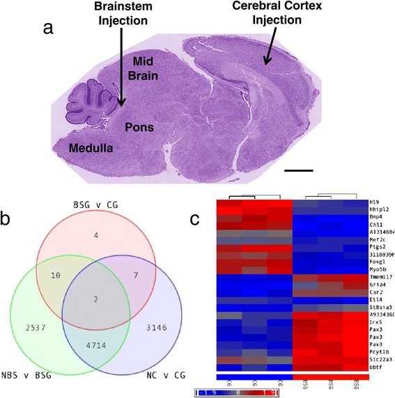
Regional differences in PDGF-B-driven mouse glioma. a Glioma was initiated in the mouse by injecting RCAS-PDGF-B virus into the brainstem or cerebral cortex of neonatal Ntv-a;Ink4a-ARF−/− mice. Shown is a sagittal section of a wildtype postnatal day 4 brain, stained with H&E, indicating the location of brainstem and cerebral cortex injections. Scale bar is 1 mM. b-c Expression profiling was conducted on the resulting Brainstem Glioma (BSG) and Cerebral Cortex Glioma (CG) and compared with age-matched normal brainstem (NBS) and normal cerebral cortex (NC), n = 3 for each. b Venn diagram showing the intersection of genes differentially expressed between NBS and BSG (green circle), between NC and CG (blue circle), and between BSG and CG (red circle). c Hierarchical clustering of 23 genes differentially regulated between BSG and CG. FDR-adjusted p-value <0.05 and fold-change ≥2.0.