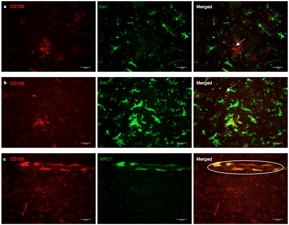
Figure 4

Double immunofluorescence of CD163 with microglia/macrophage markers Iba1 and MRC1. (a and b) Double immunofluorescence for CD163 (red) and Iba1 (green) in the occipital cortex of AD cases. Not all CD163 immunoreactive microglia stained for Iba1 (arrows), a marker highly specific for microglia. This suggests that CD163 immunoreactive parenchyma microglia might originate from systemic cells that have yet to obtain an Iba1 immunoreactive profile. The presence of microglia immunoreactive for both Iba1 and CD163 also indicate that resident microglia might be able to upregulate CD163 with stimulation from the periphery. (c) Double immunofluorescence for CD163 (red) and MRC1 (green) in the occipital cortex of an AD case. CD163 co-stains PVM with MRC1. MRC1 immunoreactivity is limited to PVM (circled). This is in concordance with findings that MRC1 is restricted to PVM despite a clear BBB breakdown.